Development and Characterization of Iron fMRI
We performed the first series of measurements using iron-oxide contrast agents for dynamic imaging of brain function, and we described theoretical and experimental comparisons with conventional fMRI signal. Relative to methods existing at the time, this new approach exhibited a large increase in detection power, which we quantified in terms of the dependencies on magnetic field strength, contrast agent dose, stimulation frequency, and tissue blood volume fraction. This technique, which we later deemed “IRON” fMRI for distinction from other evolving CBV-based methods, largely has replaced the conventional BOLD method for most preclinical fMRI applications in academic neuroscience and pharmaceutical companies. I wrote a review of the method in an anniversary issue of NeuroImage, “20 Years of functional MRI: the science and the stories.”
publications
Dynamic functional imaging of relative cerebral blood volume during rat forepaw stimulation
Dynamic measurements of regional changes in cerebral blood volume (CBV) were performed in rat models of hypercarbia and focal neuronal activation using T2-weighted imaging after injection of an intravascular contrast agent with a very long blood half-life. Calculated percent CBV change during hypercarbia was consistent with literature results from other non-invasive modalities. Equivalent percent CBV increases were found using spin- and gradient-echo images, suggesting proportional changes in blood volume for capillaries and small veins. During electrical stimulation of rat forepaw, focal CBV response to stimulation (24+/-4%) was significantly delayed relative to blood oxygen level dependent (BOLD) signal after both onset and cessation of stimulation. Poststimulus CBV decay was temporally consistent with the BOLD poststimulus undershoot. The use of exogenous agent increased the functional contrast-to-noise ratio relative to BOLD imaging by 5.7+/-1.3 at a magnetic field strength of 2 Tesla and 1.5+/-0.2 at 4.7 Tesla.
Vascular Filters of Functional MRI: Spatial Localization using BOLD and CBV Contrast.

The spatial distributions of functional activation of rat somatosensory cortex by forepaw stimulation were quantitatively compared using blood oxygen level dependent (BOLD) signal and signal weighted by cerebral blood volume (CBV). The BOLD contrast to noise (CNR) distribution showed a significant dorsal shift with respect to the CBV method at fields strengths of 2 T (0.69 6 0.09 mm) and 4.7 T (0.44 6 0.15 mm). These shifts were attributed to the gradient of resting state blood volume across somatosensory cortex and the different CNR characteristics of the two image methods. The underlying principles suggest that the CBV method has a more uniform sensitivity to percent changes in functional indicators (blood volume or deoxygenated hemoglobin) across regions of variable resting state CBV.
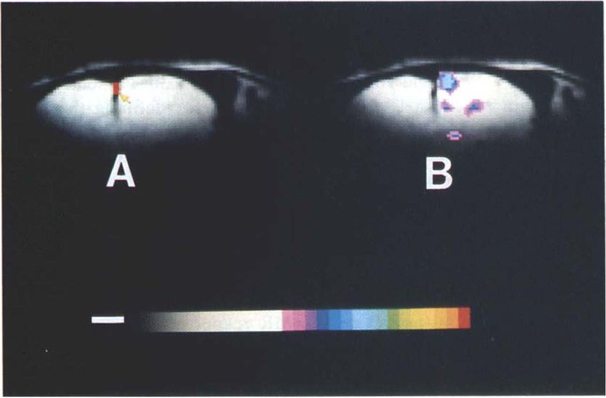
Visual motion processing investigated using contrast agent-enhanced fMRI in awake behaving monkeys.
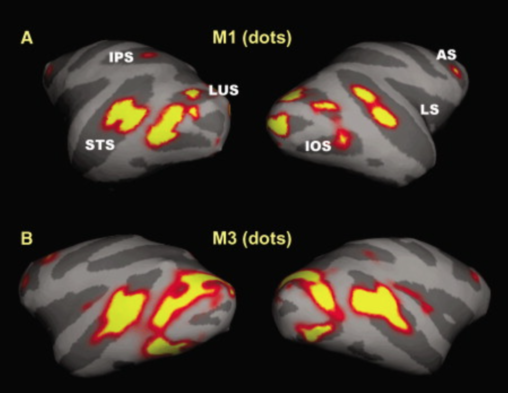
To reduce the information gap between human neuroimaging and macaque physiology and anatomy, we mapped fMRI signals produced by moving and stationary stimuli (random dots or lines) in fixating monkeys. Functional sensitivity was increased by a factor of approximately 5 relative to the BOLD technique by injecting a contrast agent (monocrystalline iron oxide nanoparticle [MION]). Areas identified as motion sensitive included V2, V3, MT/V5, vMST, FST, VIP, and FEF (with moving dots), as well as V4, TE, LIP, and PIP (with random lines). These regions sensitive for moving dots are largely in agreement with monkey single unit data and (except for V3A) with human fMRI results. Moving lines activate some regions that have not been previously implicated in motion processing. Overall, the results clarify the relationship between the motion pathway and the dorsal stream in primates.
IRON fMRI measurements of CBV and implications for BOLD signal
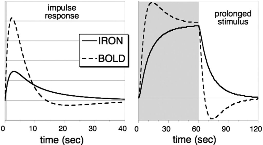
Changes in cerebral blood volume (CBV) and blood magnetization each induce changes in the transverse relaxation rate of MRI signal that are associated with changes in cerebral activity. BOLD signal, the preeminent method for non-invasive localization of task-induced brain function in human subjects, reflects a combination of changes in CBV and blood magnetization. Intravenous injection of paramagnetic contrast media, usually iron oxide particles surrounded by larger macromolecules, can overwhelm the BOLD response and sensitize signal to blood plasma volume, a method we have deemed “IRON” fMRI. The practical advantage of this technique is the ability to optimize blood magnetization at any echo time, enabling high detection power and the use of short echo times; for these reasons, IRON fMRI has become a valuable imaging tool in animal models. The temporal response of blood plasma volume is quite different from blood flow and BOLD signal; thus, CBV has been identified as a prominent source of transient features of the BOLD response. This article reviews the methodological advantages of the IRON method and how CBV measurements have informed our understanding of the BOLD response.